Weaning
The multi-disciplinary team (MDT) should be involved throughout the process of initiating weaning through to decannulation. If the patient has a neurological condition, a referral to a speech and language therapist should be made.
Criteria to commence weaning:
- The patient is able to maintain adequate gas exchange self-ventilating +/- supplemental oxygen. Occasionally patients may require non invasive ventilation (NIV) post decannulation for the management of chronic conditions such as obstructive sleep apnoea (OSA) or chronic obstructive pulmonary disease (COPD)
- There are no signs of deteriorating bronchopulmonary infection or excessive pulmonary secretions
- The patient has a stable lung status with oxygen therapy less than 40%1
- The initial reason for the insertion of the tracheostomy has been resolved and/or been considered (e.g. upper airway obstruction, cranial nerve palsy) 2,3
- The patient is cardiovascularly stable
Stages of weaning
Cuff Deflation
The cuff provides some protection from aspiration and the presence of the inflated cuff means that the patient becomes unaccustomed to managing their own secretions and swallowing. Before cuff deflation, warn the patient about the possibility of a change in airway sensation and that they may cough.
Using a synchronised suction/cuff deflation technique deflate the cuff slowly. If the patient does have difficultly with continuous coughing that does not resolve with time and reassurance and/or signs of aspiration, re-inflate the cuff and check the cuff pressure using a cuff manometer.

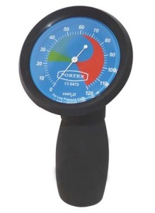
A cuff Manometer is used to measure the air pressure inside the cuff
Persistent coughing may be due to difficulties managing oral secretions due to poor swallow and/or excessive secretions with a poor cough. With the former, a referral to the speech and language therapist would be appropriate.
Throughout the cuff deflation process, monitor the patient for any signs of respiratory distress (e.g. increase in respiratory rate, heart rate, work of breathing). The time a patient spends with the cuff deflated can be increased intermittently as tolerated. The ultimate aim is to build up cuff deflation for >24 hour period and can be continued overnight4.
Gloved finger occlusion
If the patient is tolerating cuff deflation, adequate airflow around the tracheostomy tube and up into the mouth/nose needs to be established before weaning can progress any further. This is carried out by occluding the tracheostomy tube with a gloved finger and feeling for air flow from the nose/mouth. During occlusion, the patient must be monitored closely for any signs of respiratory distress, if this occurs the procedure must be stopped. Good airflow can be confirmed by auscultating over the neck above the level of the tracheostomy tube.
The presence of stridor, minimal or absent breath sounds above the level of the tracheostomy tube indicates reduced airflow around the tube. Therefore, changing the tube to a smaller size and/or fenestrated tube should be considered to optimise and proceed with weaning.
One-way speaking valve
If there is adequate airflow past the tracheostomy tube, place a one-way speaking valve over the tube opening, ensuring that the cuff is deflated. The one-way speaking valve covers the opening of the tracheostomy tube allowing air in through the valve on inspiration, but closes on expiration, allowing air past the vocal cords and out through the nose and mouth.
The patient may be able to vocalise with the one-way valve in place. Encourage vocalisation and monitor signs of difficulty managing oral secretions (e.g. wet sounding voice, difficulty clearing throat of secretions).
The length of time a patient is able to tolerate a speaking valve will vary from patient to patient and can only be gauged from observing the patient’s work of breathing. For example, a patient who has a neurological deficit presenting with swallowing difficulties may only manage a period of 2-3 minutes initially. However, a patient who has no increase in work of breathing, adequate cough and swallow may tolerate several hours on an initial trial.
With the speaking valve on, the patient is exhaling past the tracheostomy tube and through the mouth/nose. This may place a greater demand on the patient’s ventilatory reserve and the patient needs to be monitored closely for signs of respiratory distress or fatigue, which if present the trial should be stopped and the patient observed for resolution of these symptoms. The aim is to build up tolerance of using the one-way valve for more than four hours in one block, however it is not advisable to leave on overnight as secretions or sleeping position may occlude the one way valve. This process can be built up intermittently and the time increased as tolerated by the patient. It is advisable to remove the speaking valve during periods of nebulisation as the additional moisture or drugs can cause the one way valve to stick.
Decannulation cap
Once the patient is tolerating an extended period of cuff deflation and at least four hours at one time with a speaking valve in situ, a trial with the decannulation cap can be considered. This is the final stage of the weaning process and the tracheostomy tube is effectively blocked off. All the air via inspiration and expiration will be directed through the nose and mouth. The cuff must always be deflated, otherwise, the patient will be unable to breathe in or out. The aim is to build up to four hours with the decannulation cap on. This may vary with patients who have undergone head/neck surgery or who have required a tracheostomy to resolve airway obstruction where the medical staff may request a longer period of time with the deacannulation cap in place before considering decannulation. During trials with a decannulation cap the patient must be monitored for signs of respiratory fatigue or distress which if present the trial should be stopped and the patient observed for resolution of these symptoms.
Troubleshooting
- Cuff remains inflated
- Reduced space around tracheostomy tube as too large or airway obstruction present
- Poor ventilatory reserve
- Excessive secretions and/or difficulty swallowing
- Anxiety
Remove one-way valve/ decannulation cap and deflate cuff. Continue weaning with a one way valve, once the patient symptoms have resolved.
Consider changing the tube to a smaller tracheostomy tube +/_ fenestrated tube or referral to ENT to investigate airway obstruction prior to proceeding with weaning.
Build up time gradually, monitoring work of breathing
Encourage patient to cough and clear into mouth or swallow. Remove speaking valve and suction if necessary. If swallow is impaired, refer to Speech and Language Therapy
Reassure and explain procedure fully to patient
Documentation
All stages of tracheostomy weaning must be clearly documented on the weaning sheet in the tracheostomy documentation pack, which can be found in Appendix 4.
Use of fenestrated tubes
A fenestrated tracheostomy tube is a tube with one or more small holes (fenestrations) on the upper surface of the tube which allow for greater airflow through the tube into the mouth and nose and can be useful for patients who are more difficult to wean or where reducing the size of the tube may be undesirable.
The weaning and decannulation process essentially remains the same. However it is important to make sure that a fenestrated inner cannula is used during weaning, but is replaced with a non fenestrated inner cannula for suctioning or if ventilation is required.
References
- Rumbak M J, Graves A, Scott M P, Sporn, G K, Walsh F W, Anderson W, McDowell; Goldman, A (1997). Tracheostomy tube occlusion protocol predicts significant tracheal obstruction to air flow in patients requiring prolonged mechanical ventilation. Critical Care Medicine: Vol. 25 (3) pages 413-417.
- Braine M; Sweby C (2006). A systematic approach to weaning and decannulation of tracheostomy tubes. British Journal of Neuroscience Nursing, vol. 2, no. 3, pages 124 – 132.
- Kent, C (2005). Tracheostomy decannulation. Respiratory Care, vol. 50, no. 4, pages 538-541
- Thompson-Ward E, Boots R, Frisby J, Bassett L ; Timm M (1999). Evaulating suitability for tracheostomy decannulation. A Critical evaluation of two management protocols. Arch Phy Med Rehab; 80: 1101–1105.

