Diagnosis
On this page:
To diagnose and determine treatment for your particular valve disease, your doctor will obtain medical history, perform a thorough physical examination and order any or all of the following special diagnostic tests.
The tests involved in diagnosing heart valve disease include:
- ECG
- Electrocardiogram, which is tracing of the heart, mainly to show heart rate and rhythm.
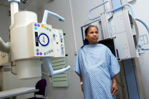
- Chest X-RAY
X-rays can provide doctors information about the size of your heart and its structure, as well as information about your lungs.
Trans-thoracic echocardiography or Echo
This is a scan of the heart, which is an ultrasound. This is non-invasive and involves a probe on the chest with some gel and shows pictures of the heart.
Trans-oesophageal echocardiography
This is an ultrasound scan of the heart. However, the probe is is placed inside the oesophagus (gullet) rather than the chest wall. This is performed without anaesthesia; however, it requires some degree of sedation where the patient becomes drowsy.
Other forms of echocardiogram
These are associated with doing some physical exercise or medication.
Computed tomography scan (CT)
A patient is placed in an x-ray tube and the heart and the rest of the body is pictured. You may need an injection in the arm, which is a form of dye, to take special pictures.
Magnetic resonance imaging scan (MRI)
A patient is placed in a tube and the heart and the rest of the body is pictured. You may need an injection in the arm, which is a form of dye, to take special pictures.
Angiography
This is to provide a picture of the coronary arteries, these are the vessels of the heart which run on the surface of the heart, and also measure other information required.
This involves an injection, sometimes in the groin and, less commonly, in an arm vessel. This is performed under local anaesthesia and takes approximately 30 minutes.