Checking for structural anomalies
On this page:
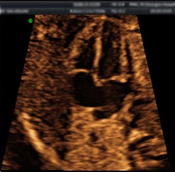
Anomaly scan
An anomaly scan is done at 21-23 weeks to check the anatomy of the baby, the baby’s growth and normal placental position. We look for normal structures in the baby’s brain, face, spine, heart, stomach, bowel, kidneys and links. If we find any structural defects we will explain these to and make an appointment for you to see a specialist to discuss it further.
Not all structural anomalies can be detected by the scan. The list below lists the chances of detection by the scan.
- Spina bifida – Open spinal defect – 90%
- Anencephaly – Defect in the formation of the skull – 99%
- Hydrocephalus – Excess fluid within the brain – 60%
- Congenital heart disease – Major heart defect – 60%
- Exomphalos /gastroschisis – Defect of the abdominal wall – 90%
- Major kidney problems – Missing or abnormal kidneys – 85%
- Major limb anomalies – Shortened or abnormally formed bones – 90%
- Diaphragmatic hernia – Defect of the muscle separating chest and abdomen – 65%
- Cleft lip and palate – 80%
- Down’s syndrome – May be associated with heart, limb, and bowel problems – 50%
- Cerebral palsy – Severe learning difficulties – not seen
- Autism – not seen – Fetal echocardiography
- Fetal echocardiography is the assessment of babies’ hearts before they are born. Most children have a normal heart; therefore, most heart scans prior to birth show no obvious anomalies.
The heart comprises four chambers, four valves, and the blood vessels bringing blood back to the heart as well as distributing blood to the lungs, body and placenta. Approximately eight in every 1,000 babies will show a heart defect. Most serious defects can be identified before babies are born when a specialised scan of the fetal heart is performed.
Although babies regularly cope with most complex anomalies prior to birth, treatment is usually required after birth (often as early as in the first few days of life) due to changes in the way blood circulates round the baby’s body following delivery.
Minor anomalies may also be detected, but aren’t always picked up by scans as minor defects rarely disturb the way the heart works. Treatment is usually unnecessary for minor anomalies. These defects happen infrequently and are usually easily dealt with, but cannot be predicted by fetal echocardiography.
For the minority of babies that show an obvious anomaly, the nature of the defect and available management options will be discussed with the parents and follow up organised as appropriate.
Fetal echocardiography is usually carried out between 20-23 weeks of gestation and a single scan is generally adequate to assess the fetal heart. However, in some situations the fetal heart can be assessed as early as 13-14 weeks of gestation and provide a lot of information. In these cases, a second fetal echo is scheduled later on in pregnancy.
Chromosomal imbalances
Chromosomal imbalances, such as Down’s Syndrome (Trisomy 21) occur in around one in every 1000 deliveries. Irrespective of what your decision would be to carry on or to terminate an affected pregnancy, you may wish to know about this before birth.
No woman is at 0% chance of carrying a baby with Down’s syndrome. The chance increases with maternal age but the vast majority of babies affected are born to younger women.
Procedures
Several non-invasive techniques have proven useful to define the chance of chromosomal imbalances during pregnancy:
- Ultrasound examination at 11-14 weeks
- Maternal biochemistry at 15-18 weeks
- Ultrasound examination at 20-23 weeks
Women booked at St George’s University Hospitals NHS Foundation Trust are all offered ultrasound examination at 11-14 weeks and 20-23 weeks as part of the package of their pregnancy care.
The fetus is seen by performing an ultrasound scan through your abdomen. However, at this early stage an internal (vaginal) scan would have to be performed to allow the fetal anatomy to be properly examined. Ultrasound scanning is an established procedure which is thought to be safe and does not harm the fetus. We will measure the fetus and look for major structural differences and also measure the amount of fluid behind the neck of the fetus.
Maternal biochemistry at 15-18 weeks is not routinely offered as part of the care package at St George’s University Hospitals NHS Foundation Trust. This procedure is regarded less accurate than the combination of the other two tests combined with maternal age. However, if you wish to have the procedure this can be arranged by the unit staff.
The result of these tests provides an individual chance, expressed as a percentage. However, these tests alone cannot tell you if your fetus has the typical number of chromosomes or not.
The test result will help us to place you in a low or a high chance group. Usually it is felt that a chance above 1/150 is sufficiently high to call for an invasive test to be offered to explore the chromosome of the fetus further.
Three invasive procedures are available to explore this, each of which carries the same risk (around 1%) of miscarriage or fetal loss:
- Chorionic villus sampling (CVS) at 11-14 weeks
- Amniocentesis from 15 weeks
- Cordocentesis from 20 weeks
It is up to you and your partner to decide whether or not you wish to risk miscarriage in order to know for certain if the baby has a chromosomal imbalance.
Personalised antenatal care of pregnancies suspected or diagnosed with Down syndrome
This document has been written for the use in the fetal medicine department at St George’s University Hospitals NHS Foundation Trust. This document offers evidence based guidance to obstetricians and midwives about the on-going management of pregnancies suspected or diagnosed with Down syndrome. There is currently no national antenatal guideline for the on-going care, and therefore guidance and care may vary depending on trust or region.